After the stethoscope, an electrocardiogram (EKG) is the most powerful tool a nurse has for knowing what is going on with a patient’s heart. Understanding how the heart works and what an EKG is measuring is your first step towards being able to accurately read an EKG and provide the physician with the information they need to help save your patient’s life.
What is Electrocardiography?
Every time a heart beats, it is depolarized to trigger a contraction. This small electrical pulse is felt throughout the body and picked up at certain points on the skin. The EKG machine is designed to pick up that activity and then display it on a graph for the medical team to see. A total of 10 electrical cables (leads) are attached to the body – 6 across the chest and 1 on each limb – in order to get the most accurate view of how the heart is beating.
The placement of the leads is important, as is the conductive gel that holds them in place. The gel aids in receiving clear transmissions, while the specific locations of each lead helps read the heart from a different angle. When the electrical pulse is traveling toward the lead, the graph will reflect an upwards line, when its traveling away from the lead, you will note a line moving downwards. This gives doctors and nurses a better view of how the heart is working.
This graph represents what one complete heartbeat should look like on an EKG for a healthy heart:
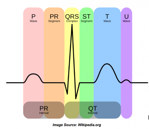
The P wave is the first electrical signal starting inside the group of cells of the Sinoatrial Node. In a normal heart, that signal will then travel through the upper chambers of the heart (atria) making them contract. That contraction pushes blood into the larger ventricles below.
The PR Interval shown on the chart is a delay in the signal that takes place at the AtrioVentricular Node. During that short delay, enough blood is emptied into the ventricles. Once the signal passes through the Bundle of His it splits, running down the left and right side bundle branches until it reaches the Purkinje fibers and turns upwards again. This is the stimulation the muscle needs to contract the heart completely and force the blood to flow out and to the rest of the body. This is what you see on the QRS stage of the graph.
That last T wave shown on the graph is the repolarization of the atria, allowing for the healthy heartbeat cycle to continue again.
What are Blocks in the EKG?
Each stage represented on the graph is a part of the normal rhythm of a heart. These stages – or blocks – can show variations on their own, and help to diagnose a cardio problem in the patient. When the interval is too long between the P and the R, the patient is said to have a first degree block. A bundle branch block is where the QRS is more than 0.12 seconds to completion. The small segments are also used to help determine the state of a heart. For example, if that smaller ST spike is continually elevated, the patient could be experiencing a myocardial infarction – or heart attack.
What Should a Nurse Know When Looking at an EKG?
Nurses who are working with cardiovascular or elderly patients may want to consider a special certification in EKG interpretation. This would put you in a position where you can take an EKG and translate the graph into readable information that can help to diagnosis a patient’s heart condition. RNs who have not received that specialized training should still be able to distinguish between a healthy and life-threatening reading, especially if they are monitoring ICU and post-surgical patients.
What is important to know for a new nurse is that a healthy heart presents a distinct pattern on an EKG. If you note anything other than this, bring it to the attention of the attending physician immediately. While not every misplaced peak is a life-threatening emergency, when it comes to your patient’s heart, it is always best to err on the side of caution.
If hearts, how they work, and their rhythms interest you, think about that certification in EKG interpretation. This not only gives you more opportunity to help cardio patients, you could be considered for higher positions in facilities and private practices that work exclusively with them.
EKGs give the medical world a unique look at the workings of the body’s most vital organ. The first step towards understanding what those peaks and valleys indicate, is knowing what the heart does, and how it is able to perform those functions.